iSpine - Spine Canal Modelling

iSpine


Spinal Imaging and Image Analysis

Mission


To provide a general method to detect different elliptical curvilinear structures under strong neighbouring disturbances.

Challenges


  1. The strong disturbance introduced by adjacent tissues and the inconsistent intensity of the spinal cord causes the detection of the spinal cord centerline and boundary to be extremely challenging tasks.
  2. Prior curvilinear structure detection approaches mainly focus on detection of circular tubular objects.
  3. Previous spinal cord detection approaches rely on extensive user inputs, training data and prior assumption of the spinal cord.[1]

Research


We propose the gradient competition anisotropy for spinal cord centerline extraction and segmentation in T1- and T2-MR images. Distinct from existing spinal cord detection approaches, the proposed method is general to the detection of different curvilinear structures, as it merely requires the minimum and maximum scales of the detection target.[1]

spinal canal modelling

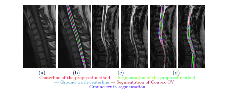
Fig. 5. Sagittal slices and corresponding segmentation results of 2 clinical cases. (a, b) The mid-sagittal slice of a T1 case. (c, d) Two sagittal slices of a T2 case.[1]

Approach


Gradient Competition Anisotropy


The propose method contains the following three components:

  1. The gradient competition descriptor sustains neighbouring disturbance to detect elliptical curvilinear objects.
    • Captures the structure orientation and curvilinearity.
  2. The orientation coherence anisotropy reliably extracts structure centerlines even if high contrast neighbouring objects are present.
    • Aids in overcoming the deterioration of the computed optimal path.
  3. The intensity coherence segmentation handles the intensity inconsistency along the structure.
    • Minimizes the intensity difference between the centerline and the voxels in vicinity of the centerline.[1]

Validation


Validation was conducted using 25 clinical datasets. Each of the three components of the proposed method was examined. Comparisons were made against five existing approaches using different criteria.[1]

Collaborators


st. joseph's hospital
lawson health research institute
robarts research
london health sciences centre
general electric
western university
digital imaging group of london