iSpine - Substructure Modelling in Spine

iSpine


Spinal Imaging and Image Analysis

Mission


To segment spinal images in a single unified framework.

Challenges


  1. Manual processing is bound to be infeasible for M³ images because of its known tediousness, inefficiency, and inconsistency.
  2. Automatic segmenting of M³ spinal images is extremely challenging in a single framework due to:
    • Entirely diverse appearances of different structures, such as vertebra and disc.
    • Completely different intensity profiles from different imaging modalities such as MRI, CT, and X-ray.
    • Substantially different shapes in varying planes, such as sagittal and axial ones, even for the same anatomic structure.
  3. Diversity of M³ images create challenges for conventional segmentation methods such as:
    • Dehydrated disc showing a dark surface and normal disc clearly showing the nucleus pulposus with stronger edges than the true boundary.
    • Completely overlapped intensity distributions of the foreground and the background of a vertebra image, which can easily confuse intensity based methods when attempting to segment both in one framework.

Research


Regression Segmentation for M³ Spinal Images


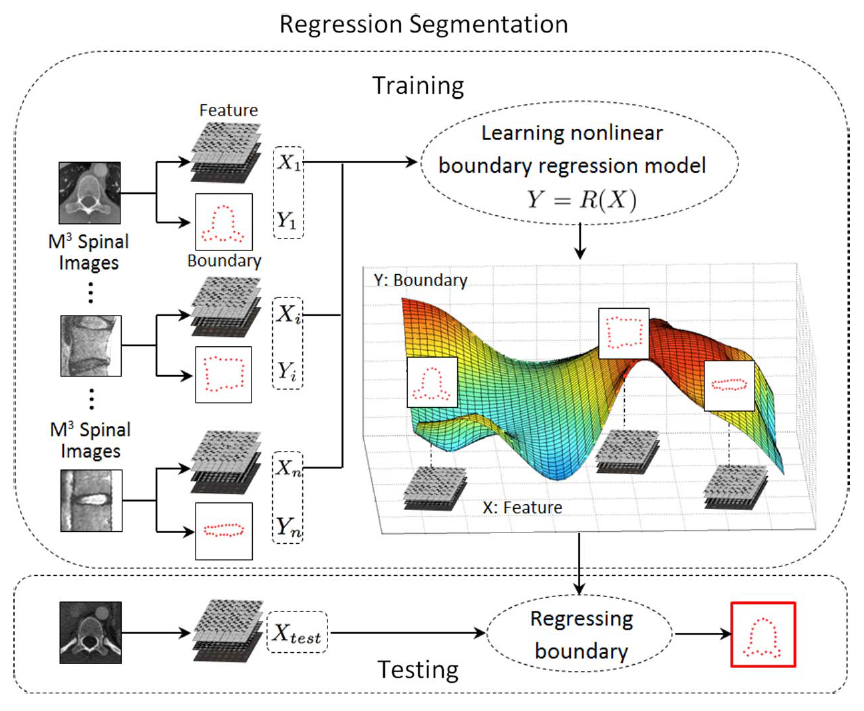
We propose a novel regression segmentation approach to successfully tackle the diversity of M³ images in a single unified framework. This approach formulates the segmentation task into a boundary regression problem that leverages the advancement of machine learning in a holistic fashion. The regression segmentation approach is fulfilled by an original multi-dimensional support vector regressor (MSVR) with a multi-kernel learning.

multiple anatomic structures

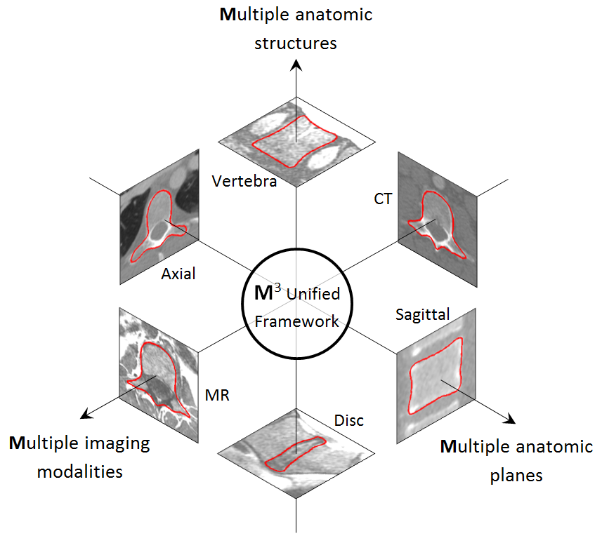
Fig. 1. Extreme diversity of spinal images including multiple anatomic structures, in multiple anatomic planes, from multiple imaging modalities. A novel approach, regression segmentation, is proposed to successfully tackle the diversity in one single unified framework. Segmentation results are shown by the solid red contours.[1]


substructure modelling


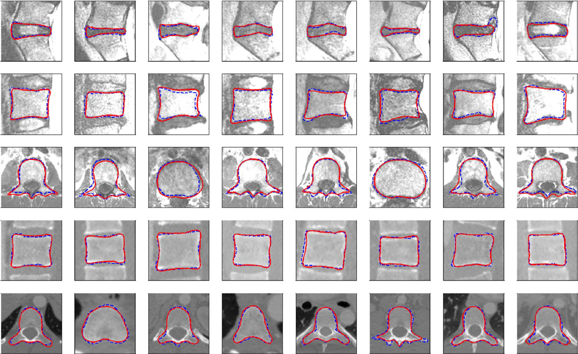
Fig. 5. Visualization of the segmentation results. Each segmentation is represented as a red solid contour, and its corresponding ground truth is represented as a blue dashed contour. Five rows respectively correspond to, sagittal disc MRI images, sagittal vertebra MRI images, axial vertebra MRI images, sagittal vertebra CT images, and axial vertebra CT images.[1]

Approach


regression segmentation

Fig. 3. Flowchart of regression segmentation framework where the segmentation task is formulated as a boundary regression problem.[1]

Training process: Learns a highly nonlinear boundary regression model.
Testing process: Predicts holistic boundary in test image based on the learned regression model.

This framework represents a boundary as the coordinates of a set of points allowing an MSVR, integrated with a multi-kernel learning technique, to fulfill the boundary regression approach. The flexible boundary representation and the highly nonlinear multi-kernel regressor are seamlessly integrated together into an effective approach for handling M³ spinal images.[1]

Validation


Validation was performed on our own and publicly available MR and CT modality datasets; containing both sagittal and axial views of lumbar and thoracic discs and vertebra structures. The strength of the regression segmentation approach was analyzed for each specific structure, modality, and plane. Comparisons between the unified M3 framework and S3 framework, regression segmentation approach versus conventional segmentation methods, multi-dimensional support vector regressor (MSVR) formulation versus single dimensional support vector regressor (SSVR) formulation, and multi-kernel MSVR versus single kernel MSVRs were conducted to validate each of the advantages.[1]

Collaborators


st. joseph's hospital
lawson health research institute
robarts research
london health sciences centre
general electric
western university
digital imaging group of london