iSpine - Stenosis

iSpine


Spinal Imaging and Image Analysis

Mission


To provide an efficient and accurate clinical tool to aid in the assessment of scoliosis.

Challenges


  1. High ambiguity and variabilty in X-rays
    • Low tissue contrast
    • Large anatomical variations[2]
    • Ambiguous boundaries[1]
  2. Inter- and intra-observer variability
  3. Multiple objects of all vertebrae[2]

Research


Structured Support Vector Regression (S2VR)


We propose the S2VR to directly predict the Cobb angles and landmarks of the spine from spinal X-ray images.

  • S2VR performs nonlinear mapping and explicit correlation modeling in a single framework.
    • Nonlinear mapping handles the relationship between the input images and high-level outputs (angles and landmarks).
    • Correlation modelling captures the correlations among outputs.
  • Formulates the prediction as a multi-output regression task.
  • Kernel target alignment is used to improve performance by increasing the discriminative ability of the kernels.[1]


BoostNet


Our proposed BoostNet performs direct spinal landmark estimation to achieve fully automatic clinical Adolescent Idiopathic Scoliosis assessment.

  • BoostNet automatically and efficiently locates spinal landmarks to provide a multi-purpose framework for robust quantitative assessment of spinal curvatures.
  • Efficiently eliminates deleterious effects of outlier features to improve robustness and generalizability.
  • Improves regression accuracy using a spinal structured multi-ouput layer.
    • Explicitly enforces dependencies between each spinal landmark to alleviate the impact of small datasets.[2]

Approach


Structured Support Vector Regression (S2VR)


The proposed framework extracts image features from spinal X-ray images and outputs coordinates, angles, and landmarks. The regression task predicts the coordinates and Cobb angles from the given input image features.[1]


structured support vector regression framework

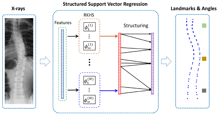
Fig. 2. The framework of the structured support vector regression (S2VR).[1]


s2vr results

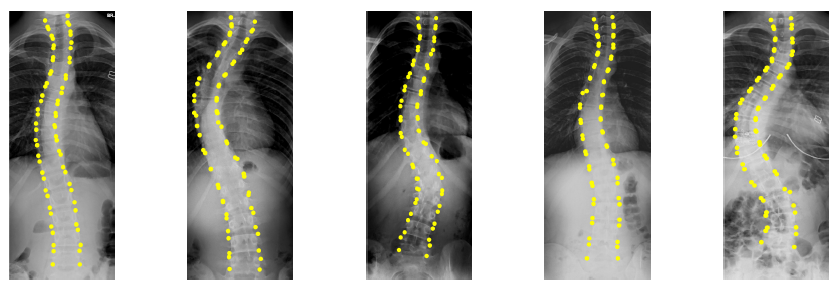
Fig. 3. Our method overcomes huge variations and high ambiguities and achieves high accuracy in landmark detection.[1]


BoostNet


The BoostNet framework consists of three parts:

  1. Series of convolutional layers to perform automated feature extraction.
  2. Newly designed BoostLayer to remove deleterious outlier features.
  3. Spinal structured multi-output layer to alleviate the impact of small datasets.[2]

boostnet framework

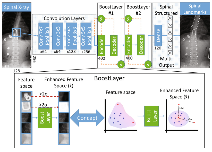
Fig. 1. Architecture of the BoostNet for landmark based AIS assessment. Relevant features are automatically extracted and any outlier features are removed by the Boost-Layer. A spinal structured multi-output layer is then applied to the output to capture the correlation between spinal landmarks.[2]


conceptual diagram of boostnet

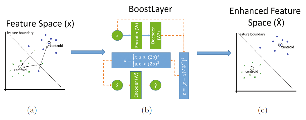
Fig. 2. Conceptualized diagram of our BoostLayer module. (a) The presence of outliers in the feature space impedes robust feature embedding. (b) The BoostLayer module detects outlier features based on a statistical properties. We use an orange dashed line to represent the outlier correction stage of the BoostLayer. For the sake of brevity, we did not include the biases and activation function in the diagram. (c) After correcting outliers, the intra-class feature variance is reduced, allowing for a more robust feature embedding.[2]


boostnet results

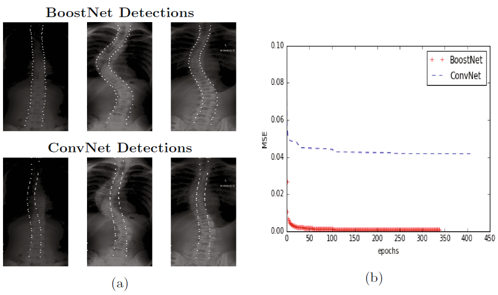
Fig. 3. Empirical results of our BoostNet algorithm. (a) The landmarks detected by our BoostNet conforms to the spinal shape more closely compared to the ConvNet detections. (b) The BoostNet converges to a much lower error rate compared to the ConvNet.[2]

Validation


S2VR was validated on a dataset composed of 439 samples from various subjects. Seventeen vertebrae from the thoracic and lumbar spine were selected for spinal shape characterization. Each vertebra is located by four landmarks with respect to four corners thus resulting in 68 points per spinal image. The leave-one-out cross-validation scheme was employed to evaluate the model's performance.[1]

BoostNet validation was conducted on a dataset consisting of 481 spinal anterior-posterior x-ray images. All images used for training and testing showed signs of scoliosis to varying extents. Seventeen vertebrae from the thoracic and lumbar spine were selected for spinal shape characterization. Each vertebra is located by four landmarks with respect to four corners thus resulting in 68 points per spinal image. The mean squared error and pearson correlation coefficients were employed to evaluate the accuracy of the model's estimations.[2]

Collaborators


st. joseph's hospital
lawson health research institute
robarts research
london health sciences centre
general electric
western university
digital imaging group of london