“`html
How to Scout Photography Locations
Scouting the perfect location can make a significant impact on the success of your photo shoots. By understanding key environments, lighting, and settings, you can create compelling images that capture the essence of your subject. This article provides a comprehensive guide for photographers aiming to scout effectively, ensuring you choose locations that enhance your creativity. We’ll explore what to look for when scouting, how to use natural landscapes to your advantage, and the importance of light and sky. Additionally, if you’re pressed for time, we’ve included quick tips and examples of ideal places to jumpstart your next session. Whether you’re a seasoned photographer or just starting, these insights promise to uplift your photography game.
3 things I’m looking for when location scouting
As every photographer knows, the right location can transform an ordinary photo into an extraordinary masterpiece. When I’m location scouting, three critical elements guide my decision: character, accessibility, and versatility. Character refers to the unique traits of a location that tell a story or complement the subject of the shoot. It might be the rustic charm of an abandoned building or the serene tranquility of a lakeside setting.
Accessibility is another vital factor. The perfect spot should be easy to reach without putting unnecessary strain on your equipment or models. An ideal location balances adventure and practicality, allowing you to concentrate fully on capturing the shot rather than worrying about logistical challenges. Finally, versatility plays a crucial role. A location should offer a range of backdrops and lighting conditions to maximize creative opportunities, keeping your shoot dynamic and engaging whilst preventing monotonous results.
Look for public land when location scouting
Exploring public lands can be a game-changer when scouting photography locations. Parks, beaches, and national reserves offer vast expanses of natural beauty with minimal restrictions, ensuring you have creative freedom to experiment with various angles and compositions. By choosing public spaces, you also avoid the complexities and costs involved with acquiring permits often necessary for private property use.
When planning a shoot in public areas, it’s essential to research and respect any local regulations or guidelines, such as time of use, group size limits, or areas designated for conservation. Public lands often provide diverse environments, from forests to fields, allowing for flexible shooting options and the spontaneous discovery of unexpected vistas. This exploration can result in fresh, authentic images that captivate your audience.
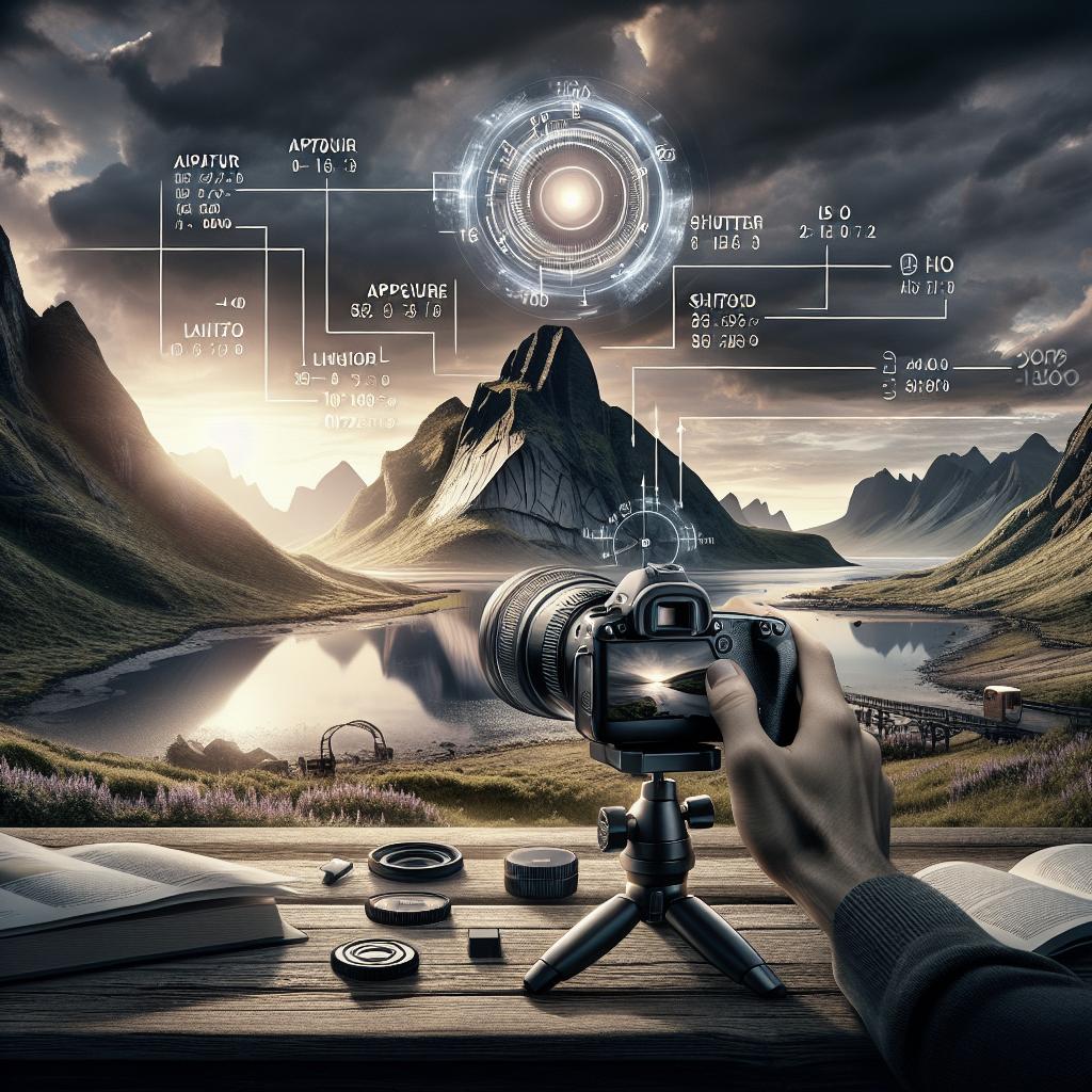
Is the sun in the right spot for sunset?
Lighting is one of the most crucial elements of photography, and the sun’s position can make or break your shoot. When planning outdoor sessions, keen photographers consider the golden hour — the time shortly after sunrise or before sunset. During these periods, the sun casts a soft, diffused light that minimizes harsh shadows and imparts a warm tone to the photos, adding a magical glow.
To ensure the sun is in the right spot for sunset shoots, tools like The Photographer’s Ephemeris can be invaluable. These apps track the solar path, helping you anticipate exactly where the sun will set in relation to your chosen location. This foresight allows for meticulous planning, ensuring you capture stunning backlit shots, silhouettes, or the captivating drama of twilight skies.
Is there a good amount of open sky at the location?
An unobstructed sky can dramatically enhance the quality and mood of outdoor photographs. A broad expanse of open sky offers ample natural light and allows photographers to frame shots creatively. A clear sky can serve as a blank canvas, highlighting the subject, while a sky filled with clouds can add texture and drama to your composition.
Alongside considering the sky, assess potential atmospheric influences at your location, such as weather and pollution, which might affect visibility and the quality of light. Planning for open skies fosters diverse photo opportunities, such as capturing the gentle hues of a sunrise or the fiery spectacle of a storm sweeping across the landscape, each evoking different emotions and telling unique stories.
Here are a few additional tips to help you find the perfect location for your next photo session:
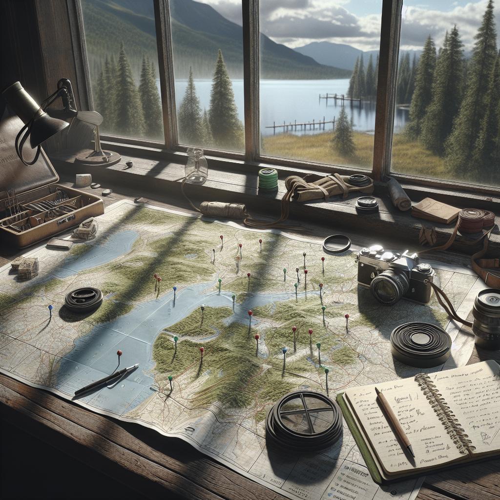
Leverage technology to streamline your location scouting process. Apps like Google Earth and Instagram geotags can offer fresh perspectives and reveal hidden gems — places discovered by other photographers that aren’t in mainstream travel guides. Social media can equally serve as an inspiration pool, showcasing popular and trending photography spots.
Consider visiting your prospective location ahead of the shoot. A pre-visit helps familiarize with the setting, evaluate potential challenges, and identify the best angles. This reconnaissance allows you to anticipate lighting conditions and prepare accordingly. Collaboration with local scouts or photographers can also yield insights into locations otherwise inaccessible or unknown.
Some examples of great locations for photography:
National parks like Yellowstone or Yosemite offer diverse landscapes that serve as exceptional backgrounds for various photography genres. From majestic waterfalls to expanse green valleys, they provide both dramatic and serene settings. Urban explorers might seek out cities like New York, which with its unique blend of architectural designs and street life, offers non-stop visual narratives.
Moreover, coastal towns and beaches introduce endless opportunities with changing tides, dynamic skies, and dramatic rock formations. Destinations such as Santorini or the Amalfi Coast showcase stunning horizons perfect for both portrait and landscape photography. The key is to remember that great locations don’t necessarily mean grand or famous ones; even your local park during foggy mornings can yield breathtaking images.
Next Steps
| Factors | Considerations |
|---|---|
| Character | Unique traits and history. |
| Accessibility | Ease of access for equipment and personnel. |
| Versatility | Diverse backdrops and lighting conditions. |
| Public Land | Free usage and diverse environments. |
| Sun’s Position | Using apps to plan for optimal light. |
| Open Sky | Maximizing natural light and atmospheric impacts. |
| Technology | Utilizing apps for discovery and planning. |
| Examples | National parks, urban settings, coastal regions. |
“`