“`html
How Filters Affect a Photograph’s Appearance
In the digital age, the use of filters in photography has become exceedingly popular. A single tap can transform an ordinary photo into something extraordinary, imbibing it with a mood or artistic flair that was perhaps not present initially. This blog post explores the multifaceted impact that filters can have on the appearance of a photograph. We will delve into the advantages, like enhanced color and sharper images, as well as the downsides, including potential image distortion and psychological effects. Ultimately, filters are powerful creative tools, but they must be used wisely to maintain photographic integrity.
Pros Of Photograph Filters
Colour Explosion
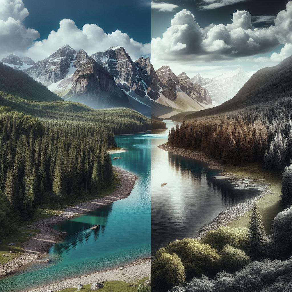
Filters can significantly enhance the colours in a photo, turning otherwise muted tones into vibrant and eye-catching hues. Many photographers turn to filters to achieve a specific mood or atmosphere, enhancing the emotional impact of the shot. For instance, a sunset photo can become even more breathtaking, with reds and oranges blazing more fiercely across the sky. This ability to amplify colours is particularly beneficial for social media platforms where “scroll-stopping” content is key.
Moreover, filters can bring out colours that might otherwise be subdued by lighting conditions or environmental factors. While natural lighting is ideal, it isn’t always available, and filters can offer a quick workaround. They allow the photographer to adjust warmth, contrast, and saturation, creating a vision that stays true to their artistic intentions.
Create Sharper Images
Filters can also play a crucial role in creating sharper images. By enhancing certain details, filters can highlight textures and patterns that lend depth and interest to an image. This can be particularly useful in landscape photography, where capturing the intricate details of nature can elevate the photograph’s overall impact.
In portrait photography, too, filters can sharpen facial features, subtly increasing contrast to emphasize the subject’s eyes, smile, or other defining characteristics. The tools that filters offer, such as clarity adjustments and edge sharpening, make it possible to rectify minor focus issues and enhance the photograph’s quality without extensive post-processing knowledge.
Get The Photograph Moving
Filters can help in creating a sense of movement in photographs, blurring the lines between static captures and kinetic stills. Motion blur filters, for example, can be used to create the illusion of speed or the passage of time, which is ideal for sports photography or artistic endeavors seeking to express dynamism.
Additionally, the creative use of filters can render still objects with implied motions, such as moving clouds over a landscape or bustling city streets. This movement enhances storytelling, inviting the viewer on a journey or to experience a moment in time as the photographer envisioned it.
Cons Of Photograph Filters
It Can Encourage Flaring
One downside of using filters is the potential for lens flaring, which can detract from image quality. Filters may inadvertently cause light to bounce around inside the lens, leading to unwanted artefacts that can ruin a photograph, particularly in bright conditions or direct light.
Photographers who shoot in challenging lighting conditions should be cautious of this risk. While some may embrace flaring for artistic purposes, it can often be mistaken for a lack of technical skill or control, making it a concern for professionals striving for crisp, clear images.
For The Professional, Filters Are Too Easy
Many professional photographers argue that relying on filters can be too easy a shortcut, bypassing fundamental photography techniques. While filters allow for quick edits, they may delay the development of essential skills such as composition, lighting, and in-camera editing.
Furthermore, filter-reliant photographers may miss out on understanding how to naturally achieve similar effects through camera settings and techniques. The art of photography heavily hinges on mastering these skills, ensuring that the photographer has complete control over the final product without needing to rely on post-processing tools.
It Can Have Negative Mental Effects
On a more profound level, the ubiquitous use of filters, particularly on social media platforms, can lead to negative mental effects. Filters often create unrealistic standards of beauty and perfection that can pressure individuals to conform, detracting from self-esteem and authentic representation.
This constant comparison with filtered, “perfect” images can contribute to anxiety and self-doubt, particularly among impressionable audiences. It is essential for both creators and consumers to be mindful of this, promoting digital authenticity and remembering that filters should emphasize, not overshadow, genuine moments and stories.
The All In All
Filters, when used deliberately, can be an incredible asset to photographers, enhancing colors, sharpening images, and even creating a sense of movement. However, caution is warranted, as over-reliance can lead to unwanted effects like flaring, diminish essential photography skills, and contribute to unrealistic imagery. By using filters judiciously, photographers can retain the integrity of their work while exploring creative horizons.
| Aspect | Pros | Cons |
|---|---|---|
| Colour Enhancement | Vibrant, eye-catching photos | Possible unrealistic representation |
| Sharpness | Enhances details and textures | Neglect of skill development |
| Movement Creation | Dynamic and engaging images | Unwanted artefacts like lens flaring |
| Psychological Impact | Powerful storytelling | Can enforce unrealistic standards |
“`