iSpine - Home Page

iSpine


Spinal Imaging and Image Analysis

Find Out More

Spine Activities


Published Books

First Spine Books 2015

Spine Web

First Spine Sharing Program!

Workshops

Annual Workshop and Challenge

Special Issues

Special Issues in the Most Prestigous Journal!

spinal imaging and image analysis

Spinal Imaging and Image Analysis
by Shuo Li, Jianhua Yoa

Citation

Shuo Li, Jianhua Yao, Spine Imaging and Image Analysis, series: LNCVB, Vol. 524, p. 261, 2015 (Spineweb)

computational radiology for orthopaedic interventions

Computational Radiology for Orthopaedic Interventions
by Guoyan Zheng, Shuo Li

Citation

Guoyan Zheng, Shuo Li, Computational Radiology for Orthopaedic Interventions Springer, series: LNCVB, 2015

computational methods and clinical apps for spine imaging

Spinal Imaging and Image Analysis
by Jianhua Yoa, Tobias Klinder, Shuo Li

Citation

Jack Yao, Tobias Klinder, and Shuo Li, Computational Methods and Clinical Applications for Spine Imaging. Springer, series: LNCVB, 2014

CSI 2016 Workshop & Challenge
The 4th Workshop on Computational Methods and Clinical Applications for Spine Imaging (CSI 2016) will be held in conjunction with MICCAI 2016 in Athen, Greece. The workshop will take place on Monday, October 17, 2016.

CSI 2015 Workshop & Challenge
We are happy to announce that our workshop and challenge proposals got accepted. For the third time, we will organize the CSI Workshop & Challenge in conjunction with MICCAI to be held in Munich, Germany, October 2015.

CSI 2014 Workshop & Challenge
The 2nd Workshop on Computational Methods and Clinical Applications for Spine Imaging (CSI 2014) was held in conjunction with MICCAI 2014 in Boston. The workshop took place on Sunday, September 14 at the Harvard Medical School.
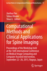
CSI 2013 Workshop
The 1st Workshop on Computational Methods and Clinical Applications for Spine Imaging (CSI 2014) was held in conjunction with MICCAI 2013 in Nagoya.

Transactions on Medical Imaging Special Issue on Spine Imaging, Image-based Modelling, and Image Guided Intervention.

Computerized Medical Imaging and Graphics Special Issue on Computational Methods and Clinical Applications for Spine Imaging.


Scope


Spinal pathologies can affect the vertebrae, intervertebral discs, ligaments, and nerve roots. These pathologies include spinal degeneration, spinal stenosis and cancer. There is, however, a lack in quantitative measurement tools in evaluation of spinal pathologies and their impact on stability and quality of life. Widespread access to 3D imaging technologies has enabled accurate visualization of spinal structures, which in turn has led to better diagnoses of spinal pathologies and more confidence in evaluating treatment effects. Such analyses, however, have thus far been mainly qualitative or semi quantitative. Quantitative analysis of large 3D image data sets is still a time consuming task motivating robust automated methods for widespread utilization.

Magnetic Resonant Imaging (MRI) and Computed Tomography (CT) are two of the popular imaging modalities used for the diagnosis of spinal pathologies. CT img are used to visualize the bony architecture of the spine, while MR img are utilized to visualize the soft tissue structures such as tumour, intervertebral discs, and spinal cord. In many scenarios, however, both bone and soft tissue structures need to be outlined to enable confident diagnosis through visualization of the diseased sites and their arrangement relative to other structures. In spinal degeneration, for instance, it is often of interest to visualize the bone and soft tissue to be able to make an accurate diagnosis or a surgical plan. When metastatic disease is present in the spine, osteolytic tumour is best visualized with MRI and osteoblastic tumour and vertebral structure are best visualized with CT; as such, both modalities are required for accurate evaluation of the extent of the disease and treatment effects. In spinal stenosis it is important to visualize both the spinal cord and the vertebral architecture to accurately plan and perform surgery. Multimodal visualization is also beneficial in treating spinal trauma and burst fracture secondary to cancer, where visualization of all involved structures (vertebrae, intervertebral discs and spinal cord/nerve roots) is critical for successful surgical planning. Analyses of multiple imaging modalities can be a challenging task, however, as the img may cover different fields of view depending on the positioning of the patient. In these cases, the radiologist or the physician may need to switch between the two img many times to be able to fully visualize the anatomical structures of interest. This motivates the development of a fused image that will enable visualization of all anatomical structures in the spine (of both hard and soft tissues) in one image using a common coordinate system.

Big Data Cloud Computing


The exponential growth and availability of health data and the existing cloud computing systems make it possible to use big data analysis tools to develop accurate models for the digital diagnoses of spine, cardiac, and eye diseases.

Our group is working on leveraging the strength of cloud computing power to help physicians with complicated clinical issue. The main chalenges faced by our group is to analyse the huge amount of unstructred data and use it to develop accurate models for the diagnosis and prognosis of clinical diseases.

Big Data Cloud Computing is provided with the collaboration with IBM

M³ iSpine Suite


The M³ iSpine Suite is a spine images analysis technique that handles multiple anatomic structures, anatomic planes, from multiple imaging modalities. It is a novel spinal images analyzing technique that is capable of performing flexible and integrated quantitative analysis of spinal structures in clinical settings. This novel approach is for the first time able to segment M³ spinal images of multiple anatomic structures, anatomic planes, and imaging modalities in a single unified framework. It is based on a novel regression segmentation approach which formulates the segmentation task as a boundary regression problem which can successfully handle the M³ images with improved segmentation and analyses accuracy, and computational effeciency.

Related Articles

1. Yunliang Cai, Sid Osman, Manas Sharma, Mark Landis, and Shuo Li. Multi-Modality Vertebra Recognition in Arbitrary Views using 3D Deformable Hierarchical Model. IEEE Transaction on Medical Imaging (TMI). 2015. Accepted.
2. Zhijie Wang, Xiantong Zhen, KengYeow Tay, Said Osman, Walt Romano, and Shuo Li. M³ Spine Segmentation. IEEE Transaction on Medical Imaging (TMI). 2015. Accepted.

M³ iSpine Suite is developed with the collaboration with IBM

References


1. Krueger, H., Noonan, V. K., Trenaman, L. M., Joshi, P., & Rivers, C. S. The economic burden of traumatic spinal cord injury in Canada.

2. Mahabaleshwarkar, R., & Khanna, R. (2014). National hospitalization burden associated with spinal cord injuries in the United States. Spinal cord, 52(2), 139-144.

3. White, N. H., & Black, N. H. (2016). Spinal cord injury (SCI) facts and figures at a glance.

4. World Health Organization, & International Spinal Cord Society. (2013). International perspectives on spinal cord injury. World Health Organization.

5. 2014-SCI-Ontario-Annual-Report [PDF]. (2014). Sciontario.org.

Collaborators


st. joseph's hospital
lawson health research institute
robarts research
london health sciences centre
general electric
western university
digital imaging group of london